OUR LAB WEBSITE HAS MOVED!
PLEASE GO to WWW.NEUROIMAGINGLAB.ORG
TO FIND OUT MORE
Functional Magnetic Resonance Imaging (fMRI)
Q: What is fMRI?
A: Simply put, functional magnetic resonance imaging (fMRI) is a tool that uses a magnetic field and pulses of radio wave energy to take pictures of organs and structures inside the body, such as the brain. It also measures neuronal activity.
Q: Is it safe?
A: Yes, fMRI is very safe and non-invasive i.e., no medication is consumed, no injections are administered.
Q: Will fMRI cause any pain?
A: No. It will not.
Q: Is there any radiation involved?
A: No. fMRI uses a strong magnet and does not emit or involve any ionizing radiation. The energy used to make magnetic resonance measurements is far less than that used in a single X-ray, and many subjects have been safely studied using magnetic resonance techniques, both locally and internationally.
Q: What are the advantages of fMRI over methods such as X-Ray, ultrasound and CT-scan?
A: These techniques take a different approach to imaging. However, using a non-invasive method, fMRI can still provide information about bodily structures, as X-ray, ultrasound, or computed tomography (CT) scan do. fMRI can also detect anomalies that cannot be seen with other imaging methods.
Q: Is there any health risk involved if I participate in the scan?
A: The risks of MRI studies are minimal, and can be performed repeatedly on human and animal subjects with no discernable long-term adverse effects.
Q: What is the difference between MRI and fMRI?
A: This is very much a technical question. Simply put, MRI takes structural images (anatomy) of the brain, while fMRI technology uses blood flow and blood oxygen levels to take functional images (brain activity) of the brain.
Q: What measures are taken to ensure the fMRI scans conducted are safe?
A: There have been possible risks and discomforts reported, however, the lab always ensures that these risks are minimized.
i) The fMRI scanner utilizes magnetic fields generated by an extremely strong magnet and will attract magnetic metal that is brought into the scanner area. Non-magnetic metallic materials might cause localized heating or generate artifacts in the images. Hence, we apply stringent requirements that all participants have to meet before being recruited. For example, people with pacemakers, metal fragments in the eye (including tattoo with metallic base), or certain metallic implants cannot participate in the study. You will be given a checklist during our pre-screening and before entering the fMRI room so that we can be sure you do not have anything harmful in or on your body. You may check past medical records to make sure of these. It is highly encouraged for you to bring along the certificate card of your implant devices, if any.
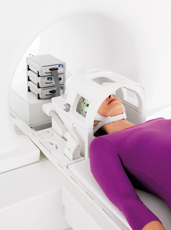
ii) In some cases, you may feel claustrophobic due to the narrow space in which you lie, even if you have never had a history of this problem. However, since the participant will have a visual screen to look at, it is unlikely that you will experience such feelings. You can gauge the size of the space from the picture above or here.
iii) The scanner emits some loud sounds while in operation. However, we provide you with earplugs to dampen the sound. Many participants have gone through the scan with the earplugs without issues with the noise.
A: Simply put, functional magnetic resonance imaging (fMRI) is a tool that uses a magnetic field and pulses of radio wave energy to take pictures of organs and structures inside the body, such as the brain. It also measures neuronal activity.
Q: Is it safe?
A: Yes, fMRI is very safe and non-invasive i.e., no medication is consumed, no injections are administered.
Q: Will fMRI cause any pain?
A: No. It will not.
Q: Is there any radiation involved?
A: No. fMRI uses a strong magnet and does not emit or involve any ionizing radiation. The energy used to make magnetic resonance measurements is far less than that used in a single X-ray, and many subjects have been safely studied using magnetic resonance techniques, both locally and internationally.
Q: What are the advantages of fMRI over methods such as X-Ray, ultrasound and CT-scan?
A: These techniques take a different approach to imaging. However, using a non-invasive method, fMRI can still provide information about bodily structures, as X-ray, ultrasound, or computed tomography (CT) scan do. fMRI can also detect anomalies that cannot be seen with other imaging methods.
Q: Is there any health risk involved if I participate in the scan?
A: The risks of MRI studies are minimal, and can be performed repeatedly on human and animal subjects with no discernable long-term adverse effects.
Q: What is the difference between MRI and fMRI?
A: This is very much a technical question. Simply put, MRI takes structural images (anatomy) of the brain, while fMRI technology uses blood flow and blood oxygen levels to take functional images (brain activity) of the brain.
Q: What measures are taken to ensure the fMRI scans conducted are safe?
A: There have been possible risks and discomforts reported, however, the lab always ensures that these risks are minimized.
i) The fMRI scanner utilizes magnetic fields generated by an extremely strong magnet and will attract magnetic metal that is brought into the scanner area. Non-magnetic metallic materials might cause localized heating or generate artifacts in the images. Hence, we apply stringent requirements that all participants have to meet before being recruited. For example, people with pacemakers, metal fragments in the eye (including tattoo with metallic base), or certain metallic implants cannot participate in the study. You will be given a checklist during our pre-screening and before entering the fMRI room so that we can be sure you do not have anything harmful in or on your body. You may check past medical records to make sure of these. It is highly encouraged for you to bring along the certificate card of your implant devices, if any.
ii) In some cases, you may feel claustrophobic due to the narrow space in which you lie, even if you have never had a history of this problem. However, since the participant will have a visual screen to look at, it is unlikely that you will experience such feelings. You can gauge the size of the space from the picture above or here.
iii) The scanner emits some loud sounds while in operation. However, we provide you with earplugs to dampen the sound. Many participants have gone through the scan with the earplugs without issues with the noise.
Our EEG-fMRI Research Study
Q: What project am I participating in when I sign up for the EEG-fMRI study?
A: The study is entitled Developmental changes in functional connectivity underlying visuospatial and emotional processing. This project involves three different age groups:
i) 12 - 17+ years old
ii) 18 - 26 years old
iii) 55 - 80 years old
Q: What are the pre-requisites of this EEG-fMRI study?
A: We require healthy chinese participants.
Q: Is this study gender-specific?
A: No. We welcome both male and female participants for this study.
Q: Is the research study being legal?
A: Yes. All our research studies are approved by Institutional Review Boards (IRB) such as NUS-IRB, SingHealth-IRB and NHG-IRB (DSRB). In this particular study, we are reviewed by the NUS-IRB.
Q: Who conducts the experiment?
A: Only trained staff are allowed to be actively involved in the experiment, following a specific set of requirements and professional protocol which included the research assistants and radiographer in Duke-NUS Medical School. They are required to pass an MRI safety test and undergo many hours of supervised training before they conduct the scans independently.
Q: Is the research study safe?
A: Yes. We use only non-invasive techniques in the study.
Q: Some studies require the participant to be injected or to take certain drugs, does this apply to this research study?
A: No. We require no contrast agents to be injected or taken orally.
Q: If I do not feel comfortable during the scan, can I pull out?
A: Yes. You are allowed to withdraw from the study at any time before or during the scan, without any consequence. You will be given an emergency bell prior to entering the scanner.
Q: Do I have to pay any fees for the scanning session?
A: No charges apply to you. The scan is done for research purposes, hence all costs are covered by the lab. Additionally, you will be reimbursed for the time you spend participating in our study.
Q: Can I be scanned if I am having fever?
A: No. Your health is our concern. Contact us a day before to get reschedule if you have a fever.
Q: I had forgotten about an important event on scan day, can I reschedule the scan?
A: Please contact us at least 4 days before the scan to reschedule. As the scanner is utilized by multiple laboratories within Duke-NUS, as well as other external collaborators, we need to ensure that scanner charges and scanner availability for other studies are being handled effectively.
Q: What will I exactly be doing for the EEG-fMRI study?
A: The flow of what participants will go through on scan day is as follows:
1) Brief on procedures & going through of MR Safety Checklist
2) Set up of EEG Cap + Concurrent completion of questionnaires
3) Practice of tasks to be performed in scanner
4) Enter MRI scanner for scan (~1.5hrs)
5) Shower
6) IQ Test
7) Reimbursement
Total time: 3.5 hours
Q: Are there any benefits for me in participating in the EEG-fMRI study?
A: There are no direct benefits for you. It is anticipated that the research findings will help us understand developmental changes in human brain functional connectivity. This could inform future methods of detecting and predicting irregular functional changes as in neurodevelopmental disorders, and developing possible targeted intervention.
Typically, an fMRI scan performed for medical purposes can cost up to $500/30 minute-scan. This study is conducted at no cost to you. You will also be compensated up to $80 or more for the 3.5 hours spent in participating in the study.
The lab also has the responsibility to inform participants of any incidental findings that we notice of their brain, if you indicate so in the MR safety form. Should these incidental findings be of concern (determined by a trained neurologist or a radiologist in our research centre), we will provide the participant with the 3D images of his/her brain such that he/she will be able to seek appropriate medical assistance.
Electroencephalography (EEG)
Q: What is EEG?
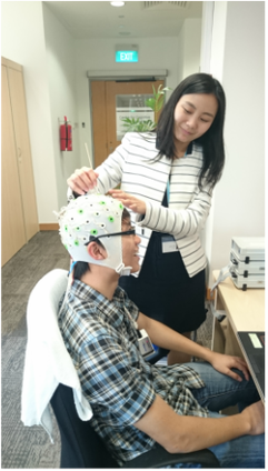
A: Electroencephalography (EEG) is a non-invasive measurement technique that records the electrical activity of the brain. you will be required to wear an EEG cap with electrodes attached on the cap and some conductance gel will be applied to your scalp for brain signal recording. You can refer to the picture over here. The gel can be washed off easily and we will provide you with the necessary toiletries, towel and hair dryer if needed.
Q: Will EEG cause any pain?
A: No. EEG is a non-invasive technique to record brain signals. The electrodes are attached on the surface of the scalp with some conductance gel applied, hence nothing will be injected into the brain nor cause pain to the participant.
Q: What is concurrent EEG-fMRI?
A: A concurrent EEG-fMRI experiment measures EEG signals of the brain while the participant is undergoing an fMRI scan. That means you will wear an EEG cap while lying in the scanner and both your brain wave patterns (EEG) and your brain activity (fMRI) will be scanned and recorded simultaneously.
Q: How many types of EEG are currently available?
A: There are 3 main types: i) Dry EEG Electrodes ii) Gel-Based EEG Electrodes for fMRI and iii) Gel-Based EEG Electrodes for MisMatch Negativity (MMN) measurement.
Q: Which type of EEG is being used in the study?
A: We only used gel-based EEG in our study. For concurrent EEG-fMRI study, we are using the gel-based EEG for fMRI. For other non-EEG-fMRI-concurrent studies, we are using MMN-EEG.
