
OUR LAB WEBSITE HAS MOVED!
PLEASE GO to WWW.NEUROIMAGINGLAB.ORG
TO FIND OUT MORE
On-going Projects
1. Developmental changes in functional connectivity underlying visuospatial and emotional processing: a current fMRI-EEG study.
The structure and function of the human brain undergo dramatic changes during development. Increasing neuroimaging evidence has characterized structural integrity and functional activation developmental trajectories. However, it remains unclear how these regions become recruited into specialized networks in the developing brain to support specific behavioral and cognitive functions. In this study, we aim to investigate functional connectivity changes underlying visuospatial processing (supported by the default mode network, DMN) and emotional processing (supported by the salience network, SN) in the healthy developing brain. In health, posterior elements of DMN participate in visuospatial processing and SN regions often activate in response to emotional stimuli. Recently, using functional magnetic resonance imaging (fMRI), we showed that divergent functional connectivity changes of DMN and SN in patients with neurodegenerative diseases corresponded well with their visuospatial and emotional dysfunctions (Zhou et al., 2010). Nevertheless, not much is known about the spatial-temporal connectivity dynamics within and between these two networks. We therefore propose employing both task-free and task-based fMRI techniques to examine the normal trajectory of functional connectivity within and between DMN and SN supporting visuospatial/emotional functions in the developing brain in vivo. We hypothesize that the within network connectivity of the DMN and SN will strengthen from childhood to adulthood, and that this will be accompanied by an increase in anti-correlation between the activity of the two networks. More importantly, we are performing concurrent fMRI-EEG recordings, integrating high temporal resolution from EEG and high spatial resolution from fMRI. This study will provide novel insights into functional brain maturation and dynamic functional connectivity, paving the way for future studies to detect and predict aberrant functional changes in neurodevelopmental disorders.
The structure and function of the human brain undergo dramatic changes during development. Increasing neuroimaging evidence has characterized structural integrity and functional activation developmental trajectories. However, it remains unclear how these regions become recruited into specialized networks in the developing brain to support specific behavioral and cognitive functions. In this study, we aim to investigate functional connectivity changes underlying visuospatial processing (supported by the default mode network, DMN) and emotional processing (supported by the salience network, SN) in the healthy developing brain. In health, posterior elements of DMN participate in visuospatial processing and SN regions often activate in response to emotional stimuli. Recently, using functional magnetic resonance imaging (fMRI), we showed that divergent functional connectivity changes of DMN and SN in patients with neurodegenerative diseases corresponded well with their visuospatial and emotional dysfunctions (Zhou et al., 2010). Nevertheless, not much is known about the spatial-temporal connectivity dynamics within and between these two networks. We therefore propose employing both task-free and task-based fMRI techniques to examine the normal trajectory of functional connectivity within and between DMN and SN supporting visuospatial/emotional functions in the developing brain in vivo. We hypothesize that the within network connectivity of the DMN and SN will strengthen from childhood to adulthood, and that this will be accompanied by an increase in anti-correlation between the activity of the two networks. More importantly, we are performing concurrent fMRI-EEG recordings, integrating high temporal resolution from EEG and high spatial resolution from fMRI. This study will provide novel insights into functional brain maturation and dynamic functional connectivity, paving the way for future studies to detect and predict aberrant functional changes in neurodevelopmental disorders.
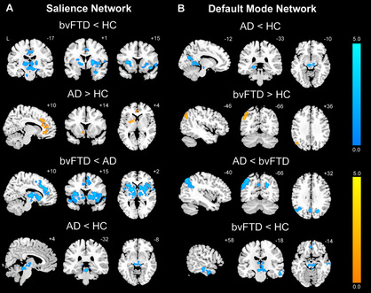
Figure 1. Divergent functional connectivity changes in AD and bvFTD (Zhou et al., Brain, 2010; Zhou et.al., Neuron, 2012)
|
|
2. Multimodal connectome analysis for differentiating subtypes within early-stage dementia and mild cognitive impairment
The prevalence of neurodegenerative diseases is predicted to increase rapidly in Singapore and around the world. As disease modifying treatments emerge, early detection and reliable discrimination of dementia is critical. This project focuses on three common types of dementia: Alzheimer’s disease (AD), behavioral variant frontotemporal dementia (bvFTD), and subcortical vascular dementia (SVD). Despite recent advances in characterizing these disorders, it has been difficult to develop clinically and scientifically useful neuroimaging biomarkers for early diagnosis and accurate prognosis. Recent work from our group and others has demonstrated that multimodal connectome analysis provides a noninvasive neuroimaging method for detecting early network-based neurodegeneration (Zhou et al, 2010; Zhou et al, 2012). The connectome is a comprehensive map of neural connections in the brain. The functional connectome, derived from task-free functional magnetic resonance imaging, maps activity synchronization between different neural assemblies. The anatomical connectome, derived from diffusion tensor imaging, measures white matter pathways connecting brain regions within networks. In this proposal, we hypothesize that integrated anatomical and functional connectome assays will prove sufficiently sensitive to (1) differentiate between early-stage AD, bvFTD, and SVD; and (2) identify amnestic and non-amnestic mild cognitive impairment (MCI) subjects who are at high risk for conversion to dementia. Moreover, we will investigate the relationships between genetic risk factors and connectome assays in mild dementia and prodromal stages. In collaboration with clinicians from National Neuroscience Institute of Singapore and Singapore General Hospital, these studies are the first step toward multimodal neuroimaging assays, seeking knowledge that will translate into clinical and scientifically useful biomarkers for early diagnosis and treatment of dementia.
The prevalence of neurodegenerative diseases is predicted to increase rapidly in Singapore and around the world. As disease modifying treatments emerge, early detection and reliable discrimination of dementia is critical. This project focuses on three common types of dementia: Alzheimer’s disease (AD), behavioral variant frontotemporal dementia (bvFTD), and subcortical vascular dementia (SVD). Despite recent advances in characterizing these disorders, it has been difficult to develop clinically and scientifically useful neuroimaging biomarkers for early diagnosis and accurate prognosis. Recent work from our group and others has demonstrated that multimodal connectome analysis provides a noninvasive neuroimaging method for detecting early network-based neurodegeneration (Zhou et al, 2010; Zhou et al, 2012). The connectome is a comprehensive map of neural connections in the brain. The functional connectome, derived from task-free functional magnetic resonance imaging, maps activity synchronization between different neural assemblies. The anatomical connectome, derived from diffusion tensor imaging, measures white matter pathways connecting brain regions within networks. In this proposal, we hypothesize that integrated anatomical and functional connectome assays will prove sufficiently sensitive to (1) differentiate between early-stage AD, bvFTD, and SVD; and (2) identify amnestic and non-amnestic mild cognitive impairment (MCI) subjects who are at high risk for conversion to dementia. Moreover, we will investigate the relationships between genetic risk factors and connectome assays in mild dementia and prodromal stages. In collaboration with clinicians from National Neuroscience Institute of Singapore and Singapore General Hospital, these studies are the first step toward multimodal neuroimaging assays, seeking knowledge that will translate into clinical and scientifically useful biomarkers for early diagnosis and treatment of dementia.
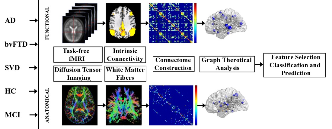
Multimodal connectome analysis
3. Brain structural and functional changes underlying cognitive decline in healthy aging
Decline in speed of processing (SOP) is a highly robust feature of cognitive aging and has been targeted for intervention by cognitive training. Deterioration of white matter (WM) integrity and loss of grey matter (GM) volume have been found associated with decreased SOP in healthy aging (Chee 2009; Kennedy 2009). However, it is still largely unknown to what extent age-related structural brain changes contribute to age-associated cognitive decline (Fjell 2010). In collaboration with CNL led by Prof. Chee, this project examines brain structural and functional changes (fMRI/DTI) of aging and their implications for understanding age-related decline in cognition (SOP) in both cross-sectional and longitudinal manners. Novel approaches to analyze longitudinal data will be developed to explore the interactions between brain and behavioral measures. The resulting complex age-dependent brain effects on cognitive performance would provide new insights into the neurobiological mechanism of age-related cognitive decline.
Decline in speed of processing (SOP) is a highly robust feature of cognitive aging and has been targeted for intervention by cognitive training. Deterioration of white matter (WM) integrity and loss of grey matter (GM) volume have been found associated with decreased SOP in healthy aging (Chee 2009; Kennedy 2009). However, it is still largely unknown to what extent age-related structural brain changes contribute to age-associated cognitive decline (Fjell 2010). In collaboration with CNL led by Prof. Chee, this project examines brain structural and functional changes (fMRI/DTI) of aging and their implications for understanding age-related decline in cognition (SOP) in both cross-sectional and longitudinal manners. Novel approaches to analyze longitudinal data will be developed to explore the interactions between brain and behavioral measures. The resulting complex age-dependent brain effects on cognitive performance would provide new insights into the neurobiological mechanism of age-related cognitive decline.
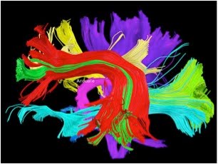
Figure 2. DTI fiber tracking (Brain Connectivity Challenge 2009)
|
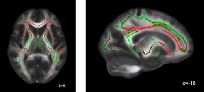
Figure 3. White matter clusters (red) where FA is significant negatively related with age (corrected p <0.05)
|
4. Anatomical and functional connectome changes in persons at risk for psychosis
The neurodevelopment model of schizophrenia predicts that individuals with an At-Risk Mental State (ARMS) will display cognitive and physiological abnormalities before the appearance of full-blown symptoms (Murray 2004; Pflueger 2007). Altered brain connectivity has been hypothesized as a major mechanism underlying cognitive deficits in schizophrenia or persons at-risk for psychosis (Andreasen 1999; Camchong 2011). However, how functional and structural connectivity are disrupted in at-risk individuals remains largely unknown. In collaboration with IMH and CNL, in this project, we hypothesized that the structural and functional connectivity in the salience network (SN) would be disrupted in ARMS subjects compared to healthy controls and that these would correlate with clinical severity. The findings may facilitate early detection and treatment for at-risk persons.
The neurodevelopment model of schizophrenia predicts that individuals with an At-Risk Mental State (ARMS) will display cognitive and physiological abnormalities before the appearance of full-blown symptoms (Murray 2004; Pflueger 2007). Altered brain connectivity has been hypothesized as a major mechanism underlying cognitive deficits in schizophrenia or persons at-risk for psychosis (Andreasen 1999; Camchong 2011). However, how functional and structural connectivity are disrupted in at-risk individuals remains largely unknown. In collaboration with IMH and CNL, in this project, we hypothesized that the structural and functional connectivity in the salience network (SN) would be disrupted in ARMS subjects compared to healthy controls and that these would correlate with clinical severity. The findings may facilitate early detection and treatment for at-risk persons.
5. Structural and functional connectivity enhancement correlates with motor recovery after rehabilitation in stroke patients
Intrinsic connectivity networks (ICNs) derived from task-free fMRI have been employed to study functional connectome in health and disease. ICN disruptions, especially inter-hemispheric connectivity, significantly correlate with visual/motor deficits in patients with stroke(Carter, Shulman et al. 2012). This project is a collaborative effort with A*STAR Institute for Infocomm Research, National University Hospital, and Singapore General Hospital in the area of stroke rehabilitation. The team has showed that motor imaginary brain computer interface (MI-BCI) and robot-assisted upper-extremity training can improve the sensorimotor recovery progress after stroke (Ang, Guan et al. 2011). However, the relationship between ICN changes and sensorimotor recovery in stroke patients after training remains largely unknown. Here, we hypothesize that the functional connectivity to primary motor cortex were disrupted in patients with stroke compared to healthy controls. More importantly, we will examine whether changes in structural/functional connectivity correlate with motor recovery after rehabilitation. Functional ICN measures, allowing behaviour prediction, would pave the way for recovery prediction and intervention monitoring after stroke.
Intrinsic connectivity networks (ICNs) derived from task-free fMRI have been employed to study functional connectome in health and disease. ICN disruptions, especially inter-hemispheric connectivity, significantly correlate with visual/motor deficits in patients with stroke(Carter, Shulman et al. 2012). This project is a collaborative effort with A*STAR Institute for Infocomm Research, National University Hospital, and Singapore General Hospital in the area of stroke rehabilitation. The team has showed that motor imaginary brain computer interface (MI-BCI) and robot-assisted upper-extremity training can improve the sensorimotor recovery progress after stroke (Ang, Guan et al. 2011). However, the relationship between ICN changes and sensorimotor recovery in stroke patients after training remains largely unknown. Here, we hypothesize that the functional connectivity to primary motor cortex were disrupted in patients with stroke compared to healthy controls. More importantly, we will examine whether changes in structural/functional connectivity correlate with motor recovery after rehabilitation. Functional ICN measures, allowing behaviour prediction, would pave the way for recovery prediction and intervention monitoring after stroke.
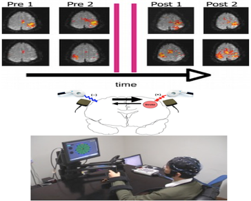
Figure 5. Combination of tDCS and MI_BCI robotic rehabilitation for stroke
6. fMRI investigation of brain-computer interface based training on selective attention and response inhibition in children with ADHD
Attention Deficit Hyperactivity Disorder (ADHD) is a developmental disorder which persists into adulthood in 50% of children. Recent evidence has cast doubts over whether present treatment with medication and behavioural management can reduce impairment in later life. The team at I2R and Duke-NUS has developed a new brain-computer interface (BCI) to treat ADHD. Our pilot studies show that it improves inattentive symptoms in children with ADHD. Elucidating the neural mechanism underlying this improvement is necessary to help us to not only gain a better picture of brain correlates related to ADHD but also allow us to identify how the intervention impacts the brain. This study aims to examine the underlying neural processes explaining its therapeutic effects. Extensive diagnostic, neuroimaging, and behavioural measures of ADHD will be assessed pre- and post-treatment during the trial. Investing modest resources in understanding how this treatment modality affects the brain will improve our understanding of the mechanisms of the treatment effect, and provide useful information if this technology can be further harnessed for future treatment of ADHD.
Attention Deficit Hyperactivity Disorder (ADHD) is a developmental disorder which persists into adulthood in 50% of children. Recent evidence has cast doubts over whether present treatment with medication and behavioural management can reduce impairment in later life. The team at I2R and Duke-NUS has developed a new brain-computer interface (BCI) to treat ADHD. Our pilot studies show that it improves inattentive symptoms in children with ADHD. Elucidating the neural mechanism underlying this improvement is necessary to help us to not only gain a better picture of brain correlates related to ADHD but also allow us to identify how the intervention impacts the brain. This study aims to examine the underlying neural processes explaining its therapeutic effects. Extensive diagnostic, neuroimaging, and behavioural measures of ADHD will be assessed pre- and post-treatment during the trial. Investing modest resources in understanding how this treatment modality affects the brain will improve our understanding of the mechanisms of the treatment effect, and provide useful information if this technology can be further harnessed for future treatment of ADHD.
